Cardiology Course
Abdominal Aortic Aneurysm Screening Exam
Why do we screen for AAA?
Many factors but predominately atherosclerotic disease can cause the wall of the aorta to weaken and bulge (aneurysm). The risk of rupture is related to the wall stress which is often estimated using the Law of Laplace. This “law” states that the stress in the AAA wall is proportional to its diameter. While this is a good rule of thumb, this may not be totally accurate. In humans, the dilation is caused by a defect in the wall and the diameter is probably not the only contributing factor to rupture. However, it is clear that the risk of rupture increases exponentially with increasing diameter, making it important to find these aneurysms early in the course so that they can be safely repaired. However, many people do not get screened even though there are very clear screening guidelines (USPSTF Aortic Aneurysm Screening Guidelines). Some studies have documented the screening rate as low as 36% of those needing a screening ultrasound exam versus patients that actually obtain one.
Please watch Abdominal Aortic Aneurysm Video below: If you need a refresher on the Abdominal Vasculature, first watch Abdominal Vasculature Anatomy.
Abdominal Vasculature Anatomy Video Abdominal Aortic Aneurysm Video
Example Images with Labels

Aorta transverse image above SMA

Aorta transverse image with SMA

Aorta transverse image above SMA

IVC long image with hepatic vein and right atrium
Heart Evaluation
Why is an ultrasound of the heart important?
Ultrasound provides an excellent way for you to understand the heart function and pathophysiology. Many of the patients you will encounter on your Adult Medicine Clinical Rotation will have pathology such as hypertension or coronary artery disease which will affect the heart. By imaging the heart, you can correlate the ultrasound findings with that of the EKG and physical exam.
What are the windows I can use to evaluate the heart?
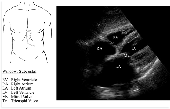
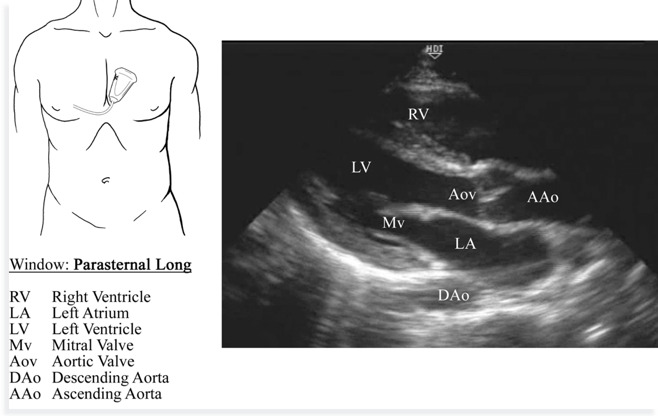
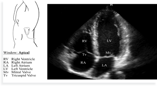
There are 3 traditional windows (views) of the heart: the subcostal, parasternal and apical windows. The key point for you is that while each of these windows are important for you to be familiar with, it is more important for you to be able to identify the structures of the heart and assess how they correlate to the EKG and physical exam findings. The key structures to identify are the left ventricle, interventricular septum, and the mitral valve. As you become comfortable assessing these structures, evaluating the relative size of the chambers and the flow across the valves using color Doppler will help you achieve a more advanced understanding of the heart.
Cardiac Windows & Structures
What can I determine about the heart function using my ultrasound images?
Determining the basic function of the heart is not as difficult as you may initially think. As long as the mitral valve is normal and the heart is in a sinus rhythm, you can evaluate the left ventricular ejection fraction (LVEF) by assessing the mitral valve opening. As can be seen in the video below, if the LVEF is normal, the mitral valve leaflet will nearly touch the interventricular septum during diastole. If it does not, then you can assume the LVEF is depressed.
Bedside Cardiac Ultrasound Video
PA Cardiology 1 Ultrasound Module and Quiz PA Cardiology 2 Ultrasound Module and Quiz