CBGM Faculty - Dr. Ashok Sharma's Lab
Bioinformatics | Big data from tiny ocular fluids | Tear Fluid | Aqueous Humor| Biomarkers| Ocular Surface

Ashok K. Sharma, PhD
Professor,
Department of Ophthalmology
Associate Director, Center for Biotechnology & Genomic Medicine
Director, CBGM Bioinformatics Core
Medical College of Georgia, Augusta University
1120 15th Street, CA-4143, Augusta, GA-30912, USA
706-721-6335
Jump to: Education Research Interests Media Articles & Publications Graduate Students & Staff
Education
- PhD, Genomic Medicine / Bioinformatics, Medical College of Georgia at Augusta University, Augusta, GA
Research Interests
With a background in computer science and a PhD in Genomic Medicine, I have experience in both the computational and biological aspects of biomedical research.
My research focus is on the development of biologically meaningful algorithms for comprehensive analysis and the visualization of high-throughput data by incorporating biological knowledge and rigorous computational methods. In the last decade, biomedical research has been revolutionized by the advent of high-throughput technologies which can generate large amounts of data. Unlocking the meaningful biological information contained inside high-throughput data is a big challenge for bench biologists. In order to facilitate the analysis of such complex data, it is crucial to develop statistical tools as well as efficient algorithms and methods to extract useful information. My primary area of research is bioinformatics, where I apply computational, mathematical, and statistical methods to solve complex biological problems. Long term goals of my lab are to solve the challenges in the analysis of data from many high-throughput technologies, especially mass spectrometry-based proteomics, and to implement these solutions in usable software tools.
Tear Fluid & Database

The images illustrates tear collection, RNA extraction and database. Tear Fluid is a thin (2-6 µm) multi-layer fluid that coats the corneal and conjunctival epithelia. The proximity of the tear Fluid to the ocular milieu, the non-invasive nature of its collection and its high protein concentration make it an attractive source for proteomic biomarker studies. We have developed a new workflow for proteomic analysis of tear fluid samples obtained with Schirmer strips that can identify over 3000 proteins in tear samples from human subjects.
We plan to collect a large number of tear samples using a standardized method and then, develop a reference tear proteomic database to prioritize, integrate, and analyze potential biomarkers. This will provide a readily accessible and permanent resource, fostering collaboration between laboratories to expand and improve the database. It is also likely that this database will help the vision research community to understand the physiological and pathological proteomic signatures in tear fluid.
Dry Eye Disease
The image illustrates how Dry Eye Disease (DED) is a complex, multifactorial condition that has become increasingly common in recent years. It is typically classified into three subtypes: aqueous deficient, evaporative, and mixed dry eye. Diagnosing DED can be challenging, as it requires both clinical expertise and an understanding of the underlying biological mechanisms. Despite advances in research, many aspects of the disease remain under active investigation. Organizations such as the Tear Fluid and Ocular Surface Society have played a key role in advancing knowledge of DED. Through their Dry Eye Workshop (DEWS) reports—most recently the DEWS III report released in 2025—they provide comprehensive insights into the causes, diagnosis, and management of the disease. Even with these efforts, important questions remain. Continued collaboration across the scientific and medical communities will be essential for developing more effective treatments and improving patient outcomes. Through the work of our lab and staff, we aim to contribute to these ongoing efforts and help advance the understanding of Dry Eye Disease.
Small non-coding RNA in tear fluid
The image illustrates the workflow for analyzing small non-coding RNA in tear fluid. The process begins with sample collection and RNA sequencing, starting with tear collection followed by RNA extraction and miRNA sequencing. Quality control is then performed through a series of steps, including adapter trimming, quality filtering, and length filtering. The processed sequences are subsequently aligned using STAR alignment. Following alignment, the RNA is annotated using Gencode, categorizing sequences into miRNA, tRNA, snRNA, and snoRNA. Finally, the data undergo analysis and are incorporated into database construction, which includes both data analysis and data visualization.

Aqueous Humor Proteomics Database
This image illustrates a step-by-step workflow for protein analysis using aqueous humor samples. The process begins with aqueous humor sample collection from the eye. The collected sample then undergoes protein extraction, where proteins are isolated from the fluid. Next, the proteins are broken down into smaller fragments through protein digestion. These resulting peptides are then cleaned and concentrated during peptide purification.
Following purification, the peptides are analyzed using LC-MS/MS (liquid chromatography–tandem mass spectrometry), which separates and identifies the peptide components. Finally, the data generated from this analysis is used for protein identification through a database search, allowing researchers to determine which proteins are present in the original sample.

Aqueous humor (AH) is the fluid in the anterior and posterior chambers of the eye that contains proteins regulating many ocular health functions, including nutrient and oxygen supply, the removal of metabolic waste, ocular immunity, and ocular shape and refraction. The dynamics of AH and the fine balance between production and drainage is essential in maintaining the physiological intraocular pressure (IOP). Therefore, identifying the protein contents of AH is vital in understanding their physiological and pathological roles in the eye.
By utilizing a large sample set, state of the art technology, and revolutionary data analysis methods, we identified the constitutive proteome of human aqueous humor, which may be useful as a reference for future studies. Discovery of AH proteomic alterations associated with glaucomatous optic neuropathy and glaucoma risk factors will help the research community at large in understanding physiological and pathological proteomics signatures in the AH.
Website: https://ahp.augusta.edu/
Bioinformatics Support
I work with an interdisciplinary team to carry out bioinformatics analysis and to pursue research projects in collaboration with other biologists. Currently, I am developing analytical methods for the large-scale measurements that are part of several Cancer and Diabetes projects. In our center (CBGM: Center for Biotechnology and Genomic Medicine), I am providing bioinformatics support for several long-term studies including The Environmental Determinants of Diabetes in the Young (TEDDY), the Phenome and Genome of Diabetes Autoimmunity (PAGODA), the Diabetic Complications Consortium (DiaComp), the Mouse Metabolic Phenotype Consortium (MMPC) and Biomarkers and Therapeutics in Cancer (BAT Cancer).
Spectrometry Analyses
- Bottom-up Proteomics
- Top-down Proteomics
- Targeted Quantification
- Post Translational Modification
- Analysis: Glycosylation and Phosphorylation
- Glycan Identification & Characterization
Vision Research Projects
- Identification of Tear Fluid Proteomic Biomarkers for Dry Eye Disease
- Proteomic Biomarkers for Glaucomatous Optic Neuropathy
- Targeting Interleukin-6 Trans-signaling in Diabetic Retinopathy
- Proteomic, Glycomic, and Glycoproteomic Profiling in Patients with Diabetic Retinopathy
- Unraveling the Role of Genetics in the Pathogenesis of Diabetic Retinopathy
Media Articles & Publications
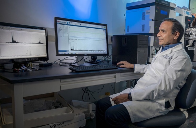
MCG scientists establish protein database to advance vision research
Scientists at the Medical College of Georgia at Augusta University have established a database that will allow researchers to better examine the underlying causes of some of the most common conditions that cause vision loss.
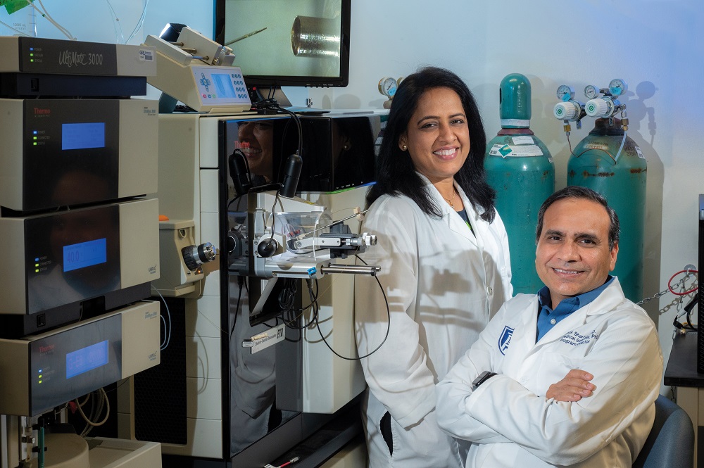
Noninvasive technique collects sufficient tear fluid to look for biomarkers of health and disease
The protective outer layer of our eyes, called the tear film, contains thousands of proteins, which provide clues about wellness and disease, and scientists have fine-tuned what they say is a non-invasive and efficient way to look at those clues.

Tear Fluid Database
The tear fluid is a thin (2-6 µm) multi-layer fluid that coats the corneal and conjunctival epithelia. Its proximity to the ocular environment, coupled with the ease of its collection and rich protein content, makes it an attractive source for proteomic biomarker studies. Novel biomarker discoveries have the potential to offer valuable insights and enhance our understanding of various ocular diseases like dry eye disease, glaucoma, and blepharitis.
A comprehensive review of tear fluid proteome alterations in dry eye disease: Insights into pathophysiology and biomarker potential
Tear fluid is a highly specialized and dynamic biofluid composed of proteins, lipids, mucins, and electrolytes that is essential for maintaining ocular surface integrity, immune defense, and tear film stability. Growing evidence from tear proteomic studieshas revealed extensive alterations in protein composition in dry eye disease (DED), reflecting a complex interplay between local ocular surface pathology and systemic inflammatory influences.
Read full story: A comprehensive review of tear fluid proteome alterations in dry eye disease: Insights into pathophysiology and biomarker potentialComprehensive profiling of the human tear fluid miRNome using small RNA sequencing
To generate a comprehensive profile of microRNAs (miRNAs) present in human tear fluid using next-generation sequencing (NGS), establish a reference miRNome for healthy human tear fluid, and investigate whether miRNA expression varies by sex, race, or age.
Read full story: Comprehensive profiling of the human tear fluid miRNome using small RNA sequencingMedical College of Georgia scientists establish protein database to advance vision research
Scientists at the Medical College of Georgia at Augusta University have established a database that will enable researchers to better study causes of vision loss.After five years of dedication and persistence, Ashok Sharma, Ph.D., associate professor and director of the bioinformatics core in the MCG Center for Biotechnology and Genomic Medicine, and a team of fellow researchers from the center, the Department of Ophthalmology, and the Culver Vision Discovery Institute have established the Aqueous Humor (AH) Proteome Database.
Read full story: Medical College of Georgia scientists establish protein database to advance vision researchNew technology enables identification of biomarkers for a wide range of diseases
Scientists have developed a way to identify biomarkers for a wide range of diseases by assessing the antibodies we are making to the complex sugars coating our cells.The new, highly sensitive Luminex Multiplex Glycan Array enables the kind of volume needed to establish associations between antibody levels in our blood to these complex sugars, or glycans, and conditions from cancer to autoimmune disease and dementia, they report in the journal Nature Communications.
Read full story: New technology enables identification of biomarkers for a wide range of diseasesGenetic discovery may help better identify children at risk for type 1 diabetes
Six novel chromosomal regions identified by scientists leading a large, prospective study of children at risk for type 1 diabetes will enable the discovery of more genes that cause the disease and more targets for treating or even preventing it.The TEDDY study’s international research team has identified the new gene regions in young people who have already developed type 1 diabetes or who have started making antibodies against their insulin-producing cells, often a precursor state to the full-blown disease that leads to a lifetime of insulin therapy.
Read full story: Genetic discovery may help better identify children at risk for type 1 diabetesPowerful anti-inflammatory molecule may block vision loss in diabetic retinopathy
A more powerful version of an anti-inflammatory molecule already circulating in our blood may help protect our vision in the face of diabetes.Diabetic retinopathy resulting from high circulating levels of glucose is the leading cause of blindness in adults. Now scientists have evidence that a man-made version of soluble gp130, or sgp130, that is 10 times more powerful than the natural one, may help avoid high levels of inflammation in the eye that occur in diabetes and avert the retinal destruction that typically follows.
Read full story: Powerful anti-inflammatory molecule may block vision loss in diabetic retinopathy
Publications
- Ahmed S, Safille S, Clifton V, Sharma S, de Paiva CS, Sharma A. A comprehensive review of tear fluid proteome alterations in dry eye disease: Insights into pathophysiology and biomarker potential. Ocul Surf. 2026 Apr;40:71-85. doi: 10.1016/j.jtos.2026.01.010. Epub 2026 Feb 3. Review. PubMed PMID: 41638552; PubMed Central PMCID: PMC13050524.
- Jones G, Mayernik D, Ahmed S, Williams E, Altman J, Lee TJ, Estes A, de Paiva CS, Martin P, Sharma S, Sharma A. Comprehensive profiling of the human tear fluid miRNome using small RNA sequencing. Ocul Surf. 2026 Apr;40:133-143. doi: 10.1016/j.jtos.2026.01.011. Epub 2026 Feb 2. PubMed PMID: 41638553; PubMed Central PMCID: PMC13050525.
- Mayernik D, Ahmed S, Williams E, Lee TJ, Estes A, Martin P, Zhi W, Jhanji V, Sharma S, Sharma A. Tear fluid database: a reference website for tear fluid proteomics. Database (Oxford). 2026 Jan 15;2026. doi: 10.1093/database/baaf091. PubMed PMID: 41540914; PubMed Central PMCID: PMC12808846.
- Ahmed S, Altman J, Jones G, Mayernik D, Williams E, Mahmoud A, Lee TJ, Zhi W, Sharma S, Sharma A. Tear fluid proteomics: a comparative study of DIA and DDA mass spectrometry. J Mass Spectrom Adv Clin Lab. 2025 Dec;38:26-36. doi: 10.1016/j.jmsacl.2025.10.001. eCollection 2025 Dec. PubMed PMID: 41158730; PubMed Central PMCID: PMC12554977.
- Williams E, Altman J, Ahmed S, Jones G, Khadanga A, Alevy D, Bollinger K, Estes A, Safille S, Lee TJ, Sharma S, Sharma A. Unveiling the levels and significance of different serpin family proteins in aqueous humor dynamics. doi: 10.1186/s12886-025-04119-3. PubMed PMID: 40389884; PubMed Central PMCID: PMC12090511.
- Lee TJ, Goyal A, Jones G, Glass J, Doshi V, Bollinger K, Ulrich L, Ahmed S, Kodeboyina SK, Estes A, Töteberg-Harms M, Zhi W, Sharma S, Sharma A. AHP DB: a reference database of proteins in the human aqueous humor. Database (Oxford). 2024 Jan 29;2024. doi: 10.1093/database/baae001. PubMed PMID: 3828493; PubMed Central PMCID: PMC10878049.
- Jones G, Altman J, Ahmed S, Lee TJ, Zhi W, Sharma S, Sharma A. Unraveling the Intraday Variations in the Tear Fluid Proteome. Invest Ophthalmol Vis Sci. 2024 Mar 5;65(3):2. doi: 10.1167/iovs.65.3.2. PubMed PMID: 38441890; PubMed Central PMCID: PMC10916888.
- Beisel A, Jones G, Glass J, Lee TJ, Töteberg-Harms M, Estes A, Ulrich L, Bollinger K, Sharma S, Sharma A. Comparative analysis of human tear fluid and aqueous humor proteomes. Ocul Surf. 2024 Mar 30;. doi: 10.1016/j.jtos.2024.03.011. [Epub ahead of print] PubMed PMID: 38561100.
- Ahmad Z, Singh S, Lee TJ, Sharma A, Lydic TA, Giri S, Kumar A. Untargeted and temporal analysis of retinal lipidome in bacterial endophthalmitis. Prostaglandins Other Lipid Mediat. 2024 Apr;171:106806. doi: 10.1016/j.prostaglandins.2023.106806. Epub 2024 Jan 5. Review. PubMed PMID: 38185280; PubMed Central PMCID: PMC10939753.
Lab Members
Graduate Students


Medical Students
Garrett N. Jones
- Medical Student (M3)
- Medical College of Georgia (MCG)
706-721-3515
Jeremy Altman
- Medical Student (M3)
- Medical College of Georgia (MCG)
706-721-3515

Sunay Kamalasanan
- Medical Student (M3)
- Medical College of Georgia (MCG)
706-721-3515


Pradyun Pulipaka
- BS/Medical Student
- Medical College of Georgia (MCG)
706-721-3515
Laboratory Faculty & Staff