Cell Imaging Core
The Cell & Tissue Imaging Core Laboratory is committed to excellence in research and education. We provide expert technical assistance and instrumentation in support of investigators.
The Core educates graduate students in the basic principles of imaging.
Publication Requirements: To cite or acknowledge the use of our core: Augusta University Medical College of Georgia Cell Imaging Core Facility, RRID:SCR_026799
Instrumentation
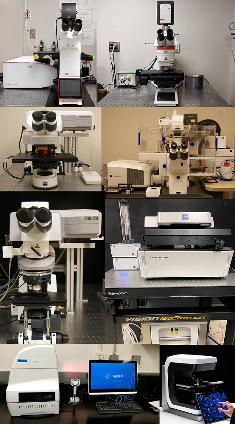
The Cell Imaging Core Laboratory has a variety of microscopes available for use.
Rules
All new users are required to be trained via the Imaging Core staff prior to using an instrument unless they can demonstrate instrument competency.
Fees
There is currently an $24.00 per hour instrument usage fee for most instruments. Utilization of these instruments is reserved for Augusta University researchers only.

The Cell and Tissue Imaging Core Laboratory has the following research capabilities:
Located in CB-2309:
- High-performance Digital Slide Scanner for Fluorescence, Brightfield and Polarization: Zeiss Axioscan 7 Digital slide scanner
- Confocal microscopy and 3D visualization: Leica Stellaris 5, Zeiss LSM780 upright and inverted confocals
- Multiphoton microscopy: Zeiss LSM780 upright multi photon, Nikon AXR upright multiphoton
- Super-resolution microscopy: Nikon STORM, Zeiss LSM780 inverted with Airyscan, Leica Stellaris 5 with Lightning
- Live imaging of cells and tissues: Zeiss Lattice Lightsheet, Leica Stellaris 5, Zeiss LSM780 inverted
- Laser capture microdissection: Leica LMD6
- Image analysis using Imaris 10.1 software (Oxford instruments)
- Measurement of oxygen consumption rate and extracellular acidification rate of live cells on a 96 well plate format: Agilent Seahorse Xfe96 Analyzer
Located within the animal facility in the Georgia Cancer Center (CN-5113):
- Live animal microscopy: Nikon AXr-Multiphoton Intravital microscope
The core is directed by Dr. Graydon Gonsalvez. Daily operations & maintenance are the responsibility of Dr. Rachel Cui.