The Mivechi Lab
Nahid F. Mivechi, PhD
Professor, Radiation Oncology
Director, Radiobiology Program
Molecular Chaperone Biology Research Group
Honors
- Distinguished Research Award, School of Graduate Studies, Medical College of Georgia, April 16, 2002
- Distinguished Faculty Award for Basic Science Research, Medical college of Georgia, May 14, 2002
- Meritorious Service Award, Department of Radiology, June 24, 2005. Augusta University Distinguished Research Award, July, 2020

Jump to: Research Summary Research Interests Selected Publications Research Team
Contact Us
The Nahid Mivechi Lab
Health Sciences Campus
GCC - M. Bert Storey Research Building
1410 Laney-Walker Blvd., CN-3156A3
(706) 721-8738
Research Summary
Dr. Nahid Mivechi has a long-standing interest in understanding the signaling pathways that govern cellular responses to disruption in protein homeostasis under physiological or pathological processes, including cancer and neurodegenerative diseases. She has extensive experience in molecular and cancer biology and has demonstrated a record of success in the research areas of regulation and function of heat shock transcription factors (HSFs) and heat shock protein (HSPs) in disease conditions.
For this research, she has developed several animal models including conventional and conditional HSF or HSP-knockout mouse lines. Her strongest contribution is in dissecting cellular and molecular mechanisms regulated by HSFs and molecular chaperone machines for cancer (Breast, T-ALL, AML, Liver), neurodegeneration (Parkinson’s disease) and metabolic diseases. A specific focus of her research program encompasses the study of the protective adaptive response of organisms to cellular and exogenous stress, which involves the activation of HSF1. Her long-standing research has established the essential function of HSF1 in hepatocellular carcinoma (HCC) development by regulating whole body metabolism (glucose utilization, gluconeogenesis, lipogenesis and cellular bioenergetics) as well as components of metabolic syndrome (insulin sensitivity and obesity). Ongoing research is devoted to study the therapeutic effects of targeting Hsf1 on HCC and establishing a possible causal relationship between Hsf1-driven alterations in hepatic or total body metabolism (obesity) and HCC development.
Research Interests
Defining the role of HSF1 in regulating heat-induced stress response in vivo.
In 2002, we reported our findings related to the effects of Hsf1 disruption in mice that we had generated. The study described two important phenomena. Firstly, using Hsf1-/- mice crossed with a Hsp70.3+/--galactosidase knock-in reporter mouse model, we demonstrated that Hsf1 is essential for the stress-induced Hsp70 expression in the whole body. However, Hsf1 does not regulate the constitutive basal expression of Hsp70 that we found to be expressed in several epithelial tissues. Secondly, we demonstrated that Hsf1 is indispensable for maintaining cellular integrity following heat stress and that cells from Hsf1-/- mice lack the ability to develop tolerance to thermal stress. Thus, the study has defined the functional significance of the Hsf1 transcriptional response, which controls the rapid induction of heat shock proteins, in the protective adaptive cellular response of organisms to cellular and environmental stress. In a more recent study, we have validated HSP70 as being an important mediator of liver cancer development. Specifically, we have discovered that genetic HSP70 inactivation impairs liver cancer initiation and progression by distinct but overlapping pathways. This includes the potentiation of carcinogen-induced DNA damage response, at the tumor initiation stage, to increase the p53-dependent surveillance response leading to the cell cycle exit or death of genomically damaged differentiated pericentral hepatocytes; these events can prevent the conversion of mature hepatocytes into more proliferating HCC progenitor cells. Subsequently activation of a MAPK/ERK negative feedback pathway diminishes oncogenic signals thereby attenuating pre-malignant cell transformation and tumor progression. Modulation of HSP70 function may be a strategy for interfering with oncogenic signals driving liver cell transformation and tumor progression, thus providing an opportunity for human cancer control.
Selected References
- Zhang Y., Serinagesh K, Dai R, Mivechi N. F. (1998). Structural Organization and promoter analysis of murine heat shock transcription factor-1 gene. J. Biol. Chem., 273:32514-32521. PMID: 9829985.
- Huang, L., Mivechi N.F, Moskofidis D. (2001). Insights into regulation and function of the major stress-induced HSP70 molecular chaperone in vivo: analysis of mice with targeted gene disruption of hsp70.1 or 70.3 genes. Cell. Biol. 21:8571-8591. PMID: 11713291
- Zhang Y., Huang L, Zhang J., Moskofidis D., Mivechi N.F. (2002). Targeted disruption of hsf1 leads to lack of thermotolerance and defines tissue-specific regulation for stress-inducible Hsp molecular chaperones. Cell. Biochemistry 86:376-393. PMID: 12112007.
- Cho W-H, Jin X, Pang J, Wang Y, Mivechi, N.F*, and Moskophidis D*. (2019). The Molecular Chaperone HSP70 Controls Liver Cancer Initiation and Progression by Regulating Adaptive DNA-Damage and MAPK/ERK Signaling Pathways (*corresponding authors). Cell. Biol. 39: e00391-18. PMID:30745413
Regulation and function of mammalian Hsf1, Hsf2 and Hsf4 in animal models of human diseases.
HSF1 is phosphorylated by multiple protein kinases that regulate its activity. In this specific area of research, we have reported that Hsf1 activity is suppressed by the ERK/MAPK signaling pathway. However, the contribution of phosphorylation in regulation of the HSF1 activity in vivo has remained elusive. To approach this issue, we generated a knock-in mouse model in which S307 and S303 have been substituted with alanine. It was predicted that these modifications might confer HSF1 to be constitutively active. In fact, our recent report confirmed that a tight posttranslational modification program via phosphorylation events on these sites is involved to regulate at the organismal level HSF1 activity and adjust age-dependent metabolic homeostasis under normal physiological conditions. Thus, these findings highlight the importance of a posttranslational mechanism (through phosphorylation at S303 and S307 sites) of regulation of the HSF1-mediated transcriptional program that moderates the severity of nutrient-induced metabolic diseases. In addition, we carried out in vivo studies to define the contribution of heat shock factor binding protein 1 (HSPBP1) in regulating HSF1 activity. HSBP1, originally isolated by Dr. R. Morimoto’s laboratory in the mid 1990s, has been proposed to repress HSF1 activity in cell lines. However, its function in vivo has remained largely elusive. To approach this issue, we used a conventional and conditional targeting strategy to disrupt the HSBP1 gene and generated two mouse models for this study. In a recent report, we provided important insights into the important role that HSBP1 may play in embryonic development, specifically regulating endoderm specification programs. However, the precise physiological role of HSBP1 in cellular function and disease conditions remains elusive, and this is a focus of ongoing research in my laboratory. With respect to the functional role of Hsf2 and Hsf4 in vivo, our studies revealed that HSF2 expression in stem cells plays a critical role in normal spermatogenesis, while HSF4 is essential for lens development and function.
Selected References
- Wang G., Ying Z, Jin X, Tu N, Zhang Y, Phillips M, Moskophidis D, Mivechi N. F. (2004). Essential requirement for both hsf1 and hsf2 transcriptional activity in spermatogenesis and male fertility. Genesis, 38:66-80. PMID: 14994269.
- Min J-N., Zhang Y, Moskophidis D, Mivechi N.F. (2004). Unique contribution of heat shock factor 4 in ocular lens development and fiber cell differentiation. Genesis, 40:205-217. PMID: 15593327
- Eroglu B., Min J-N, Zhang Y, Eroglu A, Mivechi N.F. (2014). An essential role for heat shock transcription factor binding protein 1 (HSBP1) during early embryonic development. Developmental Biology, 386: 448-460. PMID: 24380799
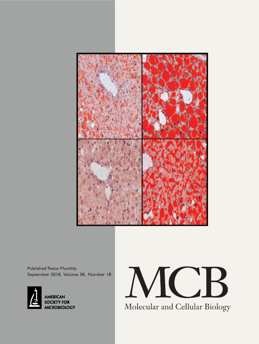
- Jin X., Qiao A, Moskophidis D, Mivechi N.F. (2018). Modulation of Heat Shock Factor 1 Activity through Silencing of Ser303/Ser307 Phosphorylation Supports a Metabolic Program Leading to Age-Related Obesity and Insulin Resistance. Molecular Cellular Biology 38: e00095-18. PMD:29941492
Function of heat shock proteins and protein phosphatase in neurodegenerative diseases
Loss of Hsp110 leads to age-dependent tau hyperphosphorylation and early accumulation of insoluble amyloid beta. An essential role of molecular chaperones in quality control is correct protein folding. Our study provides important insights into the potential function of the HSP70 machinery in tauopathy and Alzheimer’s diseases. We have demonstrated that HSP110 forms complexes with tau and protein phosphatase 2A (PP2A), and that the function of HSP110-HSP70 in these complexes is to prevent phosphorylation and aggregation of tau during aging, thus preventing cerebral plaque formation and disease progression (tauopathy). Thus, our study has provided in vivo evidence implicating the HSP70 chaperone machinery (HSP110) in the pathogenesis of Alzheimer's disease and other tauopathies. Interestingly, HSP110-deficient mice exhibit additional complex phenotypes including age-dependent development of an autoimmune hepatitis-like disease (comparable to a similar disease in human) that is associated with liver inflammation, fibrosis and sporadic liver cancer development. This phenotype of HSP110- deficient mice is currently under further investigation. Our recent work also revealed the protective role of mitochondrial dual phosphatase (Dusp) 26 in neurodegeneration and Parkinson’s disease progression
Selected References
- Eroglu B, Moskophidis D, and MivechiF. (2010). Loss of heat shock protein (Hsp) 110 leads to tau hyper-phosphorylation and early accumulation of insoluble amyloid-b peptide in mouse model of Alzheimer’s disease. Mol. Cell. Biol. 19: 4626-4643. PMID:20679486.
- Eroglu B, Kimble, D. E, Pang J, Choi J, Moskophidis D, Yanasak N, Dhandapani K.M, Mivechi N.F. (2014). Therapeutic inducers of the HSP70/HSP110 protect mice against traumatic brain injury. Journal of Neurochemistry. 130:626-641. PMID: 24903326.
- Eroglu B, Jin X, Deane S, Ozturk B, Ross O.A., Moskophidis D, and MivechiF. (2022). Dusp26 regulates mitochondrial respiration and oxidative stress and protects neuronal cell death. Cellular and Molecular Life Sciences (in Press).
Function of heat shock transcription factors in metabolic diseases and liver cancer promotion
Our research has defined an essential role for HSF1 in liver cancer promotion as well as in nutrient-induced obesity and insulin resistance. This work established the critical role of Hsf1 in metabolic reprogramming. Conceptually, we proposed a mechanistic model in which HSF1 activation promotes growth of premalignant cells and hepatocellular carcinoma (HCC) development by stimulating lipid biosynthesis and perpetuating chronic hepatic metabolic disease induced by carcinogens. Thus, genetic inactivation of Hsf1 impairs cancer progression by mitigating adverse effects of carcinogens on hepatic metabolism, liver steatosis and fibrosis as well as by preventing the development of metabolic syndrome, which includes obesity and insulin sensitivity. To further examine the function of Hsf1 in cancer development and metabolic diseases, we have generated tissue-specific Hsf1 knockout mice. Our recent work utilizing this mouse model discovered that HSF1, in addition to being key regulator of the classical chaperone response to cope with increased protein load and protein defects caused by genetic abnormalities, especially in cancer cells, also has a critical function as an information hub for anabolic metabolic and bioenergetics and protein synthesis pathways in the cell. Additional research work has discovered that specific deletion of Hsf1 from white and brown adipose tissues increases the organismal energy expenditure, regenerating the metabolic phenotype of the whole body Hsf1 mouse model. A manuscript describing this work is currently under consideration: Jin X., Qiao A, Moskophidis D, and Mivechi N.F. (2021). Heat Shock transcription factor 1 inhibits nutrient induced obesity by metabolic regulation of bioenergetics and thermogenesis in adipose tissues.
Selected References
- Jin, X., D. Moskophidis*, and N.F. Mivechi*. (2011). Heat Shock transcription factor 1 is a key determinant of HCC development by regulating hepatic steatosis and metabolic syndrome (*corresponding authors). Cell Metabolism 14:91-103. PMID: 21723507.
- Qiao, A., X. Jin, J. Pang, D. Moskophidis*, and Nahid F. Mivechi*. (2017). The transcriptional regulator of the chaperone response HSF1 controls hepatic bioenergetics and protein homeostasis (*corresponding authors). Journal Cell Biology 216: 723-741. PMID: 28183717.
Exploiting the therapeutic effects of targeting HSFs in breast cancer and in hematopoietic malignancies
A major focus of our work is to elucidate the broader contribution of HSF-driven programs in cancers of diverse types. In this context, we have shown that genetic inactivation of HSF1 suppresses the development of breast cancer induced by ErbB2/Her2 overexpression in mammary gland. Notably, the ErbB2/Neu oncogene is overexpressed in 25% of invasive/metastatic human breast cancers. Mechanistically, the inhibitory effect of HSF1 ablation on mammary cancer progression was explained through attenuation of the MAPK/ERK pathway as a consequence of the decreased levels of HSP90 chaperone protein. These results indicate a powerful tumor inhibitory pathway mediated by HSP90, which is a classical HSF1 target gene. The clinical relevance of these data is supported by recent reports indicating that high levels of HSF1 expression in the stroma is associated with poor survival prognosis. The potential contribution of HSF1 in the mammary tumor micro-environment is currently a major focus of investigation in the laboratory. This investigation uses well-established relevant mouse models.
Another major effort of our work is ongoing research on acute lymphoblastic leukemia (T-ALL) that originates from the T cell lineage. The pursuit of this research project is based on our observation that deletion of heat shock factors (HSFs) HSF1, HSF2, or HSF4 on a p53-deficient genetic background leads to significant protection against development of T-ALL. A central focus of our research effort is to explore the clinically relevant possibility that targeting HSF1 or HSF2--driven genetic and epigenetic programs that, cooperatively with oncogenes and tumor suppressor genes, drive T-ALL development and progression may be a therapeutic approach in patients. Thus, this research may provide the rationale to develop novel strategies to prevent, and perhaps treat, cancers, including T-ALL.
Selected References
- Min J., Huang L, Moskophidis D, and Mivechi N. F. (2007). Selective suppression of lymphomas by functional loss of Hsf1 in a p53-deficient mouse model for spontaneous tumors. Oncogene 26:5086-97. PMID:17310987.
- Xi C., Hu Y, Buckhaults P, Moskophidis D, and Mivechi N.F. (2012). Heat shock factor Hsf1 cooperates with ErbB2 (Her2/Neu) protein to promote mammary tumorigenesis and metastasis. J Biol. Chem. 287:35646-35657. PMCID: 22847003.
- Jin X., Eroglu B, Cho W, Yamaguchi Y, Moskophidis D, and Mivechi N.F. (2012). Inactivation of heat shock factor Hsf4 induces cellular senescence and suppresses
tumorigenesis in vivo. Mol. Cancer Res. 10:523-534. PMID: 22355043.
- Eroglu B, Pang J, Jin X, Xi C, Moskophidis D*, and Mivechi N.F.*,. (2019). HSF1-mediated control of cellular energy metabolism and mTORC1 activation drive acute T cell lymphoblastic leukemia progression (*corresponding authors). Cancer Res. 18:463-476.PMID:31744878.
Selected Publications
Program is supported by grants from the NIH/NCI
Autophagy and senescence facilitate the development of antiestrogen resistance in ER positive breast cancer. McGrath MK, Abolhassani A, Guy L, Elshazly AM, Barrett JT, Mivechi NF, Gewirtz DA, Schoenlein PV.Front Endocrinol (Lausanne). 2024 Mar 18;15:1298423. doi: 10.3389/fendo.2024.1298423. eCollection 2024. PMID: 38567308
Dusp26 phosphatase regulates mitochondrial respiration and oxidative stress and protects neuronal cell death. Eroglu B, Jin X, Deane S, Öztürk B, Ross OA, Moskophidis D, Mivechi NF.Cell Mol Life Sci. 2022 Mar 21;79(4):198. doi: 10.1007/s00018-022-04162-z.PMID: 35313355
GT198 Is a Target of Oncology Drugs and Anticancer Herbs.
Selected Publications
Amarri Johnson
Patricia Schoenlein
Jacob Chung
Sanam Kumari

Reduce the Burden
The Georgia Cancer Center at Augusta University is dedicated to reducing the burden of cancer in Georgia and across the globe through superior care, innovation, and education. Through unprecedented expansion, the Georgia Cancer Center is providing access to more first-in-the-nation clinical trials, world-renowned experts and life-saving options.
Follow the Georgia Cancer Center