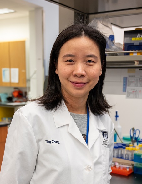
 Tianxiang Hu, PhD
Assistant Professor
Biochemistry & Molecular Biology
Georgia Cancer Center
Immunology Center of Georgia
Medical College of Georgia at Augusta University
The researches in our group focus on the investigation of molecular mechanisms regulating leukemia initiation and progression. By performing bone marrow transduction and transplantation, we have developed syngeneic mouse models of Stem Cell Leukemia/Lymphoma syndrome (SCLL) disease with mouse bone marrow cells in BALB/c mice, and human cell models with CD34+ hematopoietic stem cells in immunocompromised NSG mice. We have also established the inducible transgenic acute myeloid leukemia (AML) and Chronic myeloid leukemia (CML) mouse models through collaborations. Our previous studies deciphered the transcriptional regulatory network, including oncogenes (including cMyc, FLT3, cKit and SHP2) , Rho family GTPases, miRNAs (including miR-17/92 cluster, miR146, miR150 and miR339) and lnRNAs during leukemogenesis driven by different chromosome translocations, including BCR-FGFR1 and ZMYM2-FGFR1 in SCLL, Cbfb-MYH11 in AML, and BCR-ABL1 in CML. These research efforts on miRNAs led to the discovery of the striking contribution of IRAK1 mediated inflammatory signals in establishing immune tolerance during leukemia progression. We are now using these mouse leukemia models to investigate the underlying mechanisms in the establishment of immune evasion during leukemogenesis, with a focus on Myeloid-derived suppressor cells (MDSCs), leukemia induced neutrophils and macrophages. We hope use the newly discovered insights to direct the development of novel cancer therapy strategies.
The Tianxiang Hu Lab
Health Sciences Campus
Georgia Cancer Center - M. Bert Storey Research Building
1410 Laney Walker Blvd, Augusta, GA 30912
(706) 721-7849
Current specific research interests include:
IRAK1 regulated inflammatory signals in modulation of leukemia immune evasion.
In an investigation of the roles of miRNAs in SCLL, we identified IRAK1 as a crucial target of FGFR1 fusion kinase, and higher IRAK1 expression levels are correlated with inferior prognosis in AML, B cell lymphoma and DLBCL. Engrafted IRAK1 knockout (KO) cells did not develop leukemia in immune competent mice despite rapid leukemogenesis in immune deficient mice. T-cell depletion in immune competent mice restored leukemia development by the IRAK1KO cells, confirming a direct role of T cells in clearance of the IRAK1KO leukemia cells. In contrast, IRAK1 expressing mock control (MC) cells induce the accumulation of myeloid derived suppressor cells (MDSCs), which block the antitumor immunity of T cells, to ensure leukemia progression against immune surveillance. These observations indicate a critical on/off switch function for IRAK1 in establishing an immunosuppressive microenvironment. In addition, cytokine profiling of the MC and IRAK1KO leukemia cells identified IFNγ as a direct target of IRAK1. Engraftment using either IFNGR1-/- mice or IFNGKO leukemia cells revealed the involvement of this IRAK1 regulated IFNγ signal in modulating MDSC mediated immune suppression.
The roles of miRNA in the development of FGFR1 fusion kinase driven SCLL
miRNAs have pathogenic roles in the development of a variety of leukemias. In an effort to discover the critical roles of miRNAs in the disease progression of SCLL, we carried out a comparison of differentially expressed miRNA in normal counterparts and SCLL cells, we identified the miR-17/92 cluster and miR-339 are direct targets of FGFR1 fusion kinase. Through combined target prediction, luciferase reporter assay and western blotting, we showed that miR-17/92 promoted cell proliferation and survival by targeting CDKN1A and PTEN in B-lymphoma cell lines and primary tumors, while miR-339 promoted development of Stem Cell Leukemia/Lymphoma syndrome via downregulation of the BCL2L11 and BAX pro-apoptotic genes. The report about miR-339 is the first one to demonstrate its oncogenic effect in cancer.
FGFR1 fusion kinase mediated activation of c-Myc and Shp2 oncogenes in SCLL
In an attempt to understand how FGFR1 fusion kinase activate the expression of c-Myc oncogene, we revealed a novel functional model of FGFR1 fusion kinase, in which the full length cytoplasm-restricted chimeric kinases activated cytoplasmic signaling pathways like STAT5, to indirectly activate c-Myc expression, while a truncated form of nucleus-restricted FGFR1, generated by granzyme B cleavage of the chimeric kinases, could bind directly to MYC regulatory regions and activate its expression. In addition, we revealed the disease contribution from fusion partner gene BCR in BCR-FGFR1 driven SCLL, which is the most aggressive subtype of SCLL. With a combination of in vitro proteomic analysis with reverse phase protein array of transformed cell lines and in vivo bone marrow transduction and transplantation experiment in mouse model, we discovered that the BCR serine-threonine kinase activated the Shp2 kinase to promote leukemogenesis. This work rationalizes the necessity in developing precision therapeutics that target the consequences of the fusion partner together with the consistent aspect of the chimeric genes in chromosome translocations related leukemia.


